


Update on Implantable Sensors for Metabolic Monitoring
Progress has been made toward realizing surgically implantable electrochemical-sensor arrays.
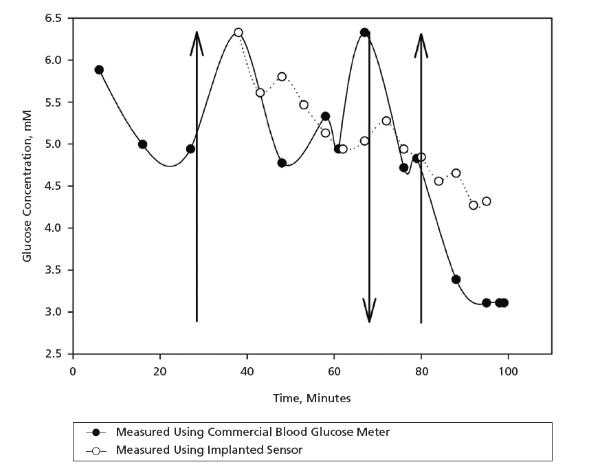
Progress has been made in a continuing effort to develop surgically implantable, biocompatible electrochemical- sensor arrays for continuous measurement of concentrations of analytes that play major roles in human and animal metabolism. This development at an earlier stage was described in "Implantable Electrochemical Sensors for Metabolic Monitoring" (ARL-0017), Defense Tech Briefs, Vol. 1, No. 4 (August 2007), page 28. To recapitulate: in the electrochemical-monitoring approach followed in this development effort, cyclic voltammetry, amperometry, squarewave voltammetry, or a combination of these techniques is used to measure the rate of catalytic oxidation of glucose by the enzyme glucose oxidase (GOX) in a reaction mediated by poly[vinyl pyridine Os(bipyridine)2Cl]- co-ethylamine (POs- EA), which is an osmium-based polycationic redox polymer. To ensure biocompatibility, the GOX is entrapped in a poly(ethylene glycol) diacrylate (PEGDA) hydrogel that has previously been demonstrated to be biocompatible.
The effort at the time of reporting the information summarized in the cited prior article was oriented particularly toward developing sensors for monitoring one analyte — glucose — to enable improved treatment of diabetic patients. At that time, it was planned to extend the effort to the fabrication and testing of sensors for monitoring lactate and pyruvate and, eventually, to implement the concept of a single array that contains sensors for monitoring glucose, lactate, and pyruvate. The progress reported since then has consisted mainly of the following:
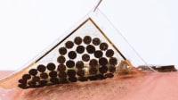
- In tests in which glucose sensors were implanted in rats, tracking of blood glucose concentrations in a limited physiological concentration range was demonstrated (see figure); and
- Lactate and pyruvate sensors were fabricated and tested in vitro.
This work was done by Michael V. Pishko of Pennsylvania State University for the U.S. Army Medical Research and Materiel Command.
This Brief includes a Technical Support Package (TSP).
Update on Implantable Sensors for Metabolic Monitoring
(reference ARL-0038) is currently available for download from the TSP library.
Don't have an account?
More From SAE Media Group
Aerospace & Defense Tech Briefs
Implantable Electrochemical Sensors for Metabolic Monitoring
Medical Design Briefs
Low-Cost Plastic Sensors Could Monitor a Range of Health Conditions
Medical Design Briefs
Wearable Sensors Printed on Natural Materials Analyze Substances Present in Sweat
Medical Design Briefs
Self-Powered Diaper Sensors Monitor Urine Sugar Levels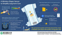
Medical Design Briefs
Wristbands Do Health Check During Workout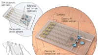
Medical Design Briefs
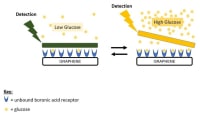
Creating Sensitive Gels for Glucose Testing
Medical Design Briefs
E-Skin Powered by Sweat Can Monitor Health
Medical Design Briefs
A Strip You Can Rely On: Tips for Designing Blood Glucose Test Strips
Aerospace & Defense Tech Briefs
NO-Generating Coats for Subcutaneous Glucose Sensors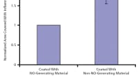
Medical Design Briefs
Paper Sensors Remove Sting of Diabetic Testing
Medical Design Briefs
Sensors Measure Uric Acid Levels
Medical Design Briefs
Bringing Glucose Monitoring to New Levels through Integrated Sensor Design
Medical Design Briefs
Skin Sensors Monitor Health
Tech Briefs
Self-Powered Paper Patch Measures Glucose
Medical Design Briefs
Wearable Sensor Measures Lactate Concentration in Real Time
Tech Briefs
Painless Paper Patch Tests Glucose Levels
Medical Design Briefs
Sensor Detects Early-Stage Parkinson’s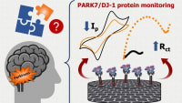
Medical Design Briefs
Flexible Sweat Sensor Based on Photonic Cellulose Nanocrystal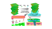
Medical Design Briefs
Hybrid Material Enables Next-Gen E-Skin
Medical Design Briefs
Tear-Based Glucose Sensor for Diabetes Monitoring
Medical Design Briefs
‘Smart’ Necklace May Track Health Through Sweat
Medical Design Briefs
Creating Cathodes for Air-Breathing Biobatteries
Medical Design Briefs
Biodegradable Implant Could Help Doctors Monitor Brain Chemistry
Medical Design Briefs
Medical Implants: Zwitterions Coating Could Prevent Blood Clotting
Medical Design Briefs
Rapid Sensor Chip for Real-Time THO Monitoring
Medical Design Briefs
Biodegradable Polymer Is Stronger and Longer Lasting
Overview
The document is a final report on the project titled "Microfabricated Multianalyte Sensor Arrays for Metabolic Monitoring," conducted by Dr. Michael V. Pishko at Pennsylvania State University, covering the period from August 15, 2004, to August 14, 2007. Funded by the U.S. Army Medical Research and Materiel Command, the project aimed to develop advanced sensor technologies for real-time metabolic monitoring, particularly focusing on glucose and lactate levels.
The report outlines the fabrication of glucose sensor arrays on gold electrodes using flexible polyimide sheets. The sensors were created through a process involving the cross-linking of glucose oxidase and redox polymers via UV-initiated free radical reactions. The methodology included conventional silicon fabrication techniques, such as mid-UV photolithography, to create five-element array microdisks. Active glucose oxidase was immobilized within photochemically polymerized hydrogels, allowing for effective entrapment on the array electrodes.
The performance of the fabricated microarray sensors was evaluated using cyclic voltammetry, demonstrating individual addressability and minimal cross-talk between adjacent elements. The sensors exhibited a linear response within the biological range, making them suitable for practical applications in metabolic monitoring. Additional electrochemical methods, including amperometry and square wave voltammetry, were employed to further assess the sensors' capabilities.
The report also discusses lactate sensors and their design, as well as the results of animal testing conducted to validate the effectiveness of the sensor arrays. Key research accomplishments and reportable outcomes are highlighted, showcasing the potential impact of these technologies on clinical and military applications.
In conclusion, the document emphasizes the significance of developing reliable, real-time monitoring systems for metabolic parameters, which can enhance patient care and operational effectiveness in military settings. The findings contribute to the broader field of biosensors and metabolic monitoring, paving the way for future research and development in this critical area. The report is approved for public release, ensuring that the knowledge gained can be shared with the scientific community and beyond.
Top Stories
NewsRF & Microwave Electronics
![]() Microvision Aquires Luminar, Plans Relationship Restoration, Multi-industry Push
Microvision Aquires Luminar, Plans Relationship Restoration, Multi-industry Push
INSIDERAerospace
![]() A Next Generation Helmet System for Navy Pilots
A Next Generation Helmet System for Navy Pilots
INSIDERDesign
![]() New Raytheon and Lockheed Martin Agreements Expand Missile Defense Production
New Raytheon and Lockheed Martin Agreements Expand Missile Defense Production
INSIDERMaterials
![]() How Airbus is Using w-DED to 3D Print Larger Titanium Airplane Parts
How Airbus is Using w-DED to 3D Print Larger Titanium Airplane Parts
NewsPower
![]() Ford Announces 48-Volt Architecture for Future Electric Truck
Ford Announces 48-Volt Architecture for Future Electric Truck
ArticlesAR/AI
Webcasts
Electronics & Computers
![]() Cooling a New Generation of Aerospace and Defense Embedded...
Cooling a New Generation of Aerospace and Defense Embedded...
Automotive
![]() Battery Abuse Testing: Pushing to Failure
Battery Abuse Testing: Pushing to Failure
Power
![]() A FREE Two-Day Event Dedicated to Connected Mobility
A FREE Two-Day Event Dedicated to Connected Mobility
Unmanned Systems
![]() Quiet, Please: NVH Improvement Opportunities in the Early Design Cycle
Quiet, Please: NVH Improvement Opportunities in the Early Design Cycle
Automotive
![]() Advantages of Smart Power Distribution Unit Design for Automotive &...
Advantages of Smart Power Distribution Unit Design for Automotive &...
Energy
![]() Sesame Solar's Nanogrid Tech Promises Major Gains in Drone Endurance
Sesame Solar's Nanogrid Tech Promises Major Gains in Drone Endurance